Retinal Diseases
The Basics
The retina is the back part of the eye responsible for providing vision. A healthy retina is essential in giving you the full visual world. When you look at an object, the image of that object is focused on to the retina, and then the visual information is passed along to the brain through the optic nerve. The macula is the central portion of the retina, responsible for the central vision. Some patients confuse the term “macula” with age-related macular degeneration, which is a common aging condition. The macula is simply the location in the retina that provides the sharp, clear central vision, such as the vision used for reading, working on the computer, seeing faces, and seeing to drive. Many conditions can affect the macula. It is therefore important to keep the macula as healthy as possible.
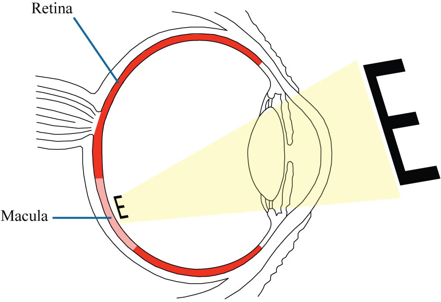
The macula is the central part of the retina. When you look at an object, the image is focused sharply on to the macula.
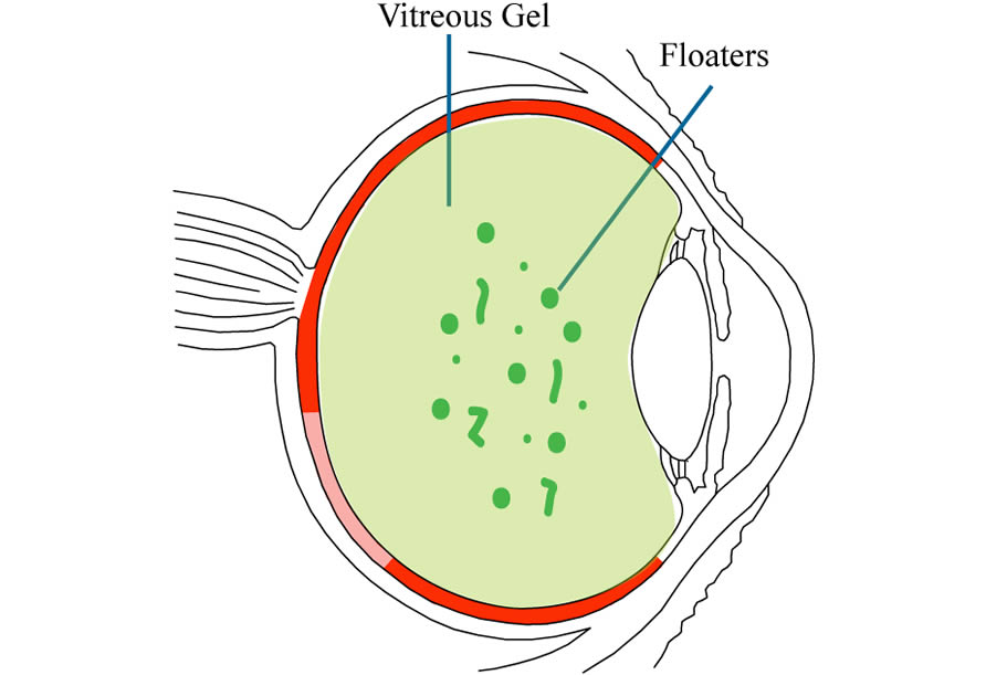
The vitreous gel starts out mostly clear. With age, the collagen fibers clump together to form floaters.
The vitreous gel is the clear gelatinous material in front of the retina. Aging of the vitreous gel is responsible for a variety of conditions in the eye, including:
- floaters
- retinal tear
- retinal detachment
- vitreous hemorrhage
- epiretinal membrane
- macular hole
Overview
Age-Related Macular Degeneration
Age-related macular degeneration (AMD) is strongly driven by a person’s age and genetic background. The majority of patients with AMD has the dry form, which tends to be associated with relatively good vision. The dry form can turn into the wet form in 20% of cases. The wet form means that there is internal fluid seepage or bleeding in the macula. The wet form is usually treated by eye injections. It is important to know that AMD primarily affects the central vision (reading, watching TV, seeing faces). The peripheral vision is usually intact even in severe cases, and this can allow many patients to enjoy many activities of daily living and often live healthy, independent lives.
Learn More About
Age-Related Macular Degeneration
Central Serous Retinopathy
Central Serous Retinopathy (CSR), also called Central Serous Chorioretinopathy (CSC), is a very common condition that affects the younger age group. It typically manifests a sudden onset of blurred vision or visual distortion in one eye without any pain. The main cause for CSR is probably anatomy. Patients with CSR often have a exuberant vasculature under the retina. This exuberant vasculature can start to leak fluid into the retina, especially when triggered by the use of corticosteroids products. The fluid in active CSR often resolves spontaneously in 3 months, which can seem like a long time if you are suffering from active CSR. Some patients can experience vision loss due to persistent fluid. A laser treatment can often be very effective in stopping the CSR fluid leakage.
Learn More About
Central Serous Retinopathy
Diabetic Retinopathy
Diabetic retinopathy is the leading cause of blindness among working-age adults. Diabetes is a disease that slowly damages the tiny blood vessels throughout your body, especially in the eyes. These blood vessels can start to leak fluid into the retina, interfering with normal vision. The most effective way to prevent vision loss in diabetes is to have regular dilated eye examinations. When a problem is discovered early, treatments can often be given to stop vision loss or help vision improve. If you have diabetes retinopathy, working closely with your diabetes doctor and your retina specialist is the best way to ensure you will have the best vision long-term. With diabetes, success comes from all the little baby steps that you take.
Learn More About
Diabetic Retinopathy
Epiretinal Membrane
Eipertinal membrane, also known as Macular Pucker, is a very common age-related condition. It is also commonly known as a “wrinkle” in the back of the eye. If the condition does not affect the vision much, it can be left alone and be monitored. If the vision becomes significantly affected, the epiretinal membrane can be removed by outpatient surgery called vitrectomy. The visual prognosis is usually good.
Learn More About
Epiretinal Membrane
Flashes & Floaters
Flashes and floaters are very common. Having them does not always mean a torn retina. On the other hand, it is definitely a good idea to see a retina specialist if you are developing any new flashes and floaters. A torn retina can usually be treated by a laser. If you notice a shadow or a shade or a thumbprint in your vision, this can mean your retina has detached, and you may need surgery to repair the retina.
Learn More About
Flashes & Floaters
Low Vision Information
Despite all the advances that modern medicine can bring, many patients still live with low vision. There is help available for these patients. Many patients with impaired vision, when given appropriate low vision aids and training, can learn to read, do work, or sometimes even drive, and so function more independently. Even patients who are legally blind can improve their quality of life by low vision services.
Learn More About
Low Vision Information
Macular Hole
Macular hole is an age-related condition that can develop in patients with age > 55. If you have a macular hole, you may notice blurred vision with one eye. If you try to cover the unaffected eye and read with the affected eye, you may notice some letters missing or distorted. Macular hole occurs because of abnormal vitreous gel aging over the macula. A macular hole can be repaired by vitrectomy, an outpatient surgical procedure.
Retinal Detachment
Retinal detachment is usually caused by abnormal aging in the vitreous gel. The vitreous gel contracts, pulls, and tears the retina. The retina then becomes dislodged from the back of the eye, resulting in a shadow or vision loss. A retinal detachment often requires surgery for repair. If you have a retinal detachment in one eye, you must have the other eye monitored in the future, since the other eye has an increased chance of developing a retinal detachment also.
Learn More About
Retinal Detachment
Retinal Vein Occlusion
Retina Vein Occlusion is a common condition. It usually develops in patients with age > 50, but it can occur in younger patients. The retina is supplied by a tree of blood vessels–arteries and veins. One of the branches of the veins can become narrowed and occluded. Sometimes the main central trunk of the vein can become occluded. When that happens, fluid can accumulate in the macula, interfering with normal vision. Treatment usually is very effective and may include eye injections and laser treatments, but they often need to be repeated.
Learn More About
Retinal Vein Occlusion
Vitreous Hemorrhage
The vitreous gel inside the eye is normally mostly clear. Some of the collagen fibers inside the vitreous gel can start to mature with age and turn into little clumps or strands, which can be visible as floaters. They are noticeable especially when looking at a bright background like a white computer screen or the blue sky. If you develop a vitreous hemorrhage, you may notice sudden appearance of more floaters in your vision. If the hemorrhage is severe, the entire vision can turn dark. There are many conditions that can cause a vitreous hemorrhage. Depending on the cause, different treatments may be needed. If you think you have develop a vitreous hemorrhage, you should see a retina specialist promptly.